
The analysis of brain wave data holds significant importance for understanding the signals sent throughout the body. By exploring this data, researchers can unravel the complexities of the human mind and develop strategies to assist individuals facing neurological challenges. In this article, we will delve into the various types of brain waves and how Python can be an effective tool for analyzing this data.
The human brain plays a pivotal role in maintaining our activity and functionality throughout life. The principles of artificial intelligence, deep learning, and artificial neural networks draw inspiration from the brain's operation. Artificial neural networks are designed to mimic the brain's neural networks to allow machines to perceive and process data similarly to humans.
This capability enables medical professionals and researchers to leverage cutting-edge technology to deduce intricate details about the brain's function and detect abnormalities related to neurological disorders. We will focus on one approach to investigate how the brain operates and analyze the signals it dispatches to various body parts.
The brain consistently produces waves of electrical activity that vary based on a person's emotional state at any given moment. Tools like EEG kits effectively capture and record these wave patterns. The programming language Python proves essential in analyzing such data, aiding in assessing a person’s alertness or level of concentration. By evaluating extensive datasets, experts can pinpoint the underlying causes of brain-related issues and explore potential treatments.
Let’s start by examining the nature of brain waves and their classifications before discussing how Python can facilitate the analysis of brain wave data.
Understanding Brain Waves
Brain waves refer to the electrical impulses utilized by neurons for intercommunication. These neurons rely on electrical signals to convey various human emotions and behaviors.
Each type of brain wave exhibits distinct frequencies based on the emotion experienced by an individual. These frequencies, measured in hertz (Hz) or cycles per second, manifest as both slow and fast waves produced by neurons. Each wave type is designated a specific name to differentiate them according to frequency.
Categories of Brain Waves
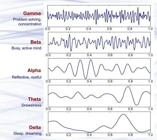
Five categories define brain waves, with delta waves being the slowest and gamma waves the fastest. The frequency of these waves influences levels of consciousness experienced by humans.
Delta Brain Waves
Delta waves are the slowest, characterized by high amplitude and a frequency between 1 and 3 Hz, typically observed during sleep.
Theta Brain Waves
These waves range in frequency from 4 to 7 Hz, occurring in a dreamy state. When frequencies are closer to the lower end, they signify a transitional phase between wakefulness and sleep, often referred to as the twilight state. Theta waves usually suggest that a person might be notably relaxed or mentally disengaged.
Alpha Brain Waves
Alpha waves function at frequencies from 8 to 12 Hz, reflecting a larger, slower waveform associated with a calm state. They emerge when an individual experiences tranquility, often after visualizing something soothing with their eyes closed.
Beta Brain Waves
Beta waves are quicker and smaller, operating within a frequency range of 13 to 38 Hz. These waves indicate heightened focus and demonstrate that an individual is fully alert and engaged in mental activities.
Gamma Brain Waves
Gamma waves are the fastest, with frequencies spanning from 38 to 42 Hz. They are less prominent than other wave types and are essential for cognitive functioning and perception. These waves are typically generated when an individual is highly alert and attuned to subtle changes in their environment.
Visualizing Various Brain Wave Forms
Capturing Brain Waves
Electroencephalography (EEG) is a widely adopted method for capturing brain waves and recording electrical activity from the scalp. This technique illustrates the overall activity of brain waves. Electrode placements on the scalp allow recordings of this activity, and EEG is generally a non-invasive method. Conversely, electrocorticography (intracranial EEG) involves an invasive approach.
This technique gauges variations in voltage from ionic current released by neurons. Clinically, EEG represents the spontaneous electrical activity recorded over time. Data collection employs multiple electrodes positioned on a subject's scalp.
Diagnosis focuses on detecting spectral content or event-related potentials over specific durations and events. The spectral content analyzes the types of neural oscillations or brain waves.
Tools for Analyzing Brain Waves
Two primary applications serve to analyze brain waves, targeting different analytical aspects.
Emotion Analysis
The brain determines human emotions, with brain waves transmitting messages associated with feelings. Analyzing these waves allows for a deeper understanding of emotional states, though interpretations can vary significantly based on cultural backgrounds and experiences. A classification algorithm is crucial for accurately interpreting the emotional dimensions of brain wave activity.
Brain-Computer Interface (BCI)
BCIs play a significant role by collecting brain wave patterns, interpreting signals, transforming them into commands, and conveying them to devices. This process operates independently of neuromuscular pathways, aiming to restore the functionality of these pathways in individuals suffering from neurological impairments.
Conditions such as cerebral palsy, stroke, or spinal cord injury can disrupt these pathways and affect the transmission of brain waves. BCIs seek to rectify such dysfunction and help individuals regain control of physical movements during their rehabilitation process.
The initial uses focused on EEG-based spelling devices and single-neuron control mechanisms. Scientists have expanded applications to include intracortical, electrocorticographic, and electroencephalographic signals, suitable for intricate control tasks. These techniques have enabled control of devices such as robotic limbs and prosthetics.
Applications of Brain-Computer Interfaces
Innovations in science and technology have popularized BCIs among researchers. These interfaces are being utilized in various applications that include:
Device Control Through Thought
Portable EEG devices, like Emotiv EPOC, capture EEG signals. The raw data undergoes denoising using the Discrete Wavelet Transform (DWT) method, while the Canonical Correlation Analysis (CCA) method extracts and categorizes the signals. Classified outcomes convert into commands that control smart devices at home.
In essence, individuals can issue commands and manage home automation devices simply by contemplating the action without needing to verbalize it.
Thought-Controlled Driving
BCIs empower individuals with physical disabilities to operate vehicles with their thoughts. Recordings of brain signals via mobile EEG devices are processed and translated into commands for action by the respective devices.
This technology acts as a reader of brain intent, translating thoughts into commands for external applications. For instance, individuals with mobility impairments can steer a wheelchair or automate car driving solely through mental focus.
Systems such as Audi AI: ME and Aicon allow passengers to manage the graphical interface through not just touch and voice, but also eye tracking, achieved through infrared sensors that detect where a user’s gaze is directed.
Neuralink: Integrating Technology within the Brain
Neuralink, co-founded by Elon Musk, is a neurotechnology firm specializing in developing brain-machine interfaces (BMIs) that can be implanted directly into the brain. These devices aim to enable the seamless transfer of brain wave signals from neurons to associated devices. The company has enlisted top neuroscientists to design an optimal BMI chip. While initial human trials were anticipated for 2020, the schedule has faced delays.
Analyzing Brain Wave Data with Python
We will now explore two methods for analyzing brain wave data using Python.
MNE Library in Python
The MNE-Python library is an open-source resource for processing, analyzing, and visualizing neuroimaging data.
Step 1: Installing Required Libraries
!pip install mne
!pip install pyvista
!pip install vtk
Step 2: Downloading and Importing Brain Wave Data for Analysis
import os
import numpy as np
import mne
# Downloading brain wave datasets
sample_data_folder = mne.datasets.sample.data_path()
sample_data_evk_file = os.path.join(sample_data_folder, ‘MEG’, ‘sample’,
‘sample_audvis-ave.fif’)
evokeds_list = mne.read_evokeds(sample_data_evk_file, baseline=(None, 0),
proj=True, verbose=False)# Show the condition names, and reassure ourselves that baseline correction has been applied. for e in evokeds_list:
print(f’Condition: {e.comment}, baseline: {e.baseline}’)
Step 3: Visualizing Various Brain Wave Data
conds = (‘aud/left’, ‘aud/right’, ‘vis/left’, ‘vis/right’)
evks = dict(zip(conds, evokeds_list))
evks[‘aud/left’].plot(exclude=[])
Output:
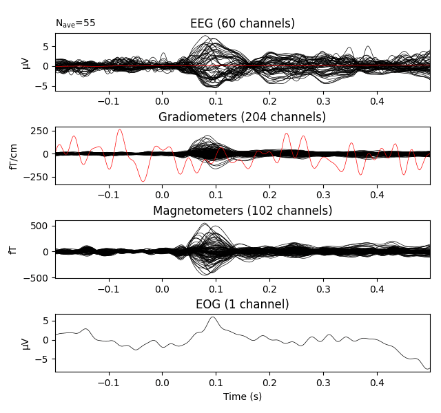
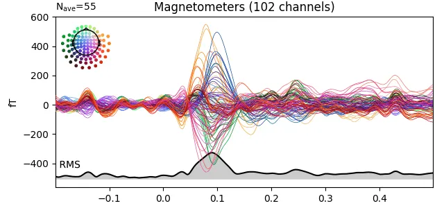
Step 4: MEG Data Visualization
Output:
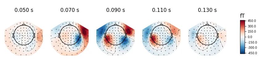
Step 5: Scalp Topography and Average Field Values
evks[‘aud/left’].plot_topomap(ch_type=’mag’, times=times, colorbar=True)
Output:
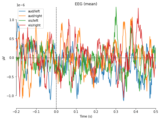
Step 6: Calculating and Visualizing Average EEG Values
return x.max(axis=1)
for combine in (‘mean’, ‘median’, ‘gfp’, custom_func):
mne. viz.plot_compare_evokeds(evks, picks=’eeg’, combine=combine)
Output:
Step 7: Additional MEG Data Visualization
linestyles=dict(left=’solid’, right=’dashed’))
temp_list = list()
for idx, _comment inenumerate((‘foo’, ‘foo’, ”, None, ‘bar’), start=1):
_evk = evokeds_list[0].copy()
_evk.comment = _comment
_evk.data *= idx # so we can tell the traces apart
temp_list.append(_evk)
mne.viz.plot_compare_evokeds(temp_list, picks=’mag’)
evks[‘vis/right’].plot_image(picks=’meg’)
mne.viz.plot_compare_evokeds(evks, picks=’eeg’, colors=dict(aud=0, vis=1),
linestyles=dict(left=’solid’, right=’dashed’),
axes=’topo’, styles=dict(aud=dict(linewidth=1),
vis=dict(linewidth=1)))
mne.viz.plot_evoked_topo(evokeds_list)
subjects_dir = os.path.join(sample_data_folder, ‘subjects’)
sample_data_trans_file = os.path.join(sample_data_folder, ‘MEG’, ‘sample’,
‘sample_audvis_raw-trans.fif’)
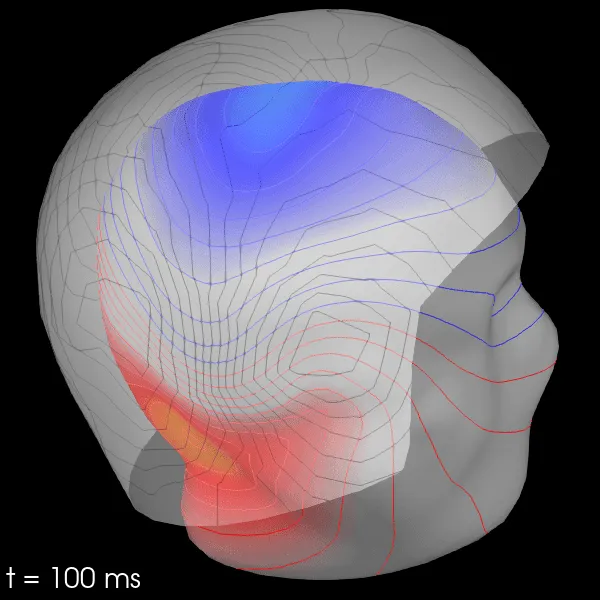
Step 8: Creation of 3D Field Maps
subject=’sample’, subjects_dir=subjects_dir)
evks[‘aud/left’].plot_field(maps, time=0.1)
Output:
Employing Deep CNN Networks
No-Sang Kwak and others have recommended utilizing the SSVEP (steady-state visual evoked potential) classifier which is based on a convolutional neural network (CNN-1). This classifier incorporates two hidden layers (with kernel sizes of 1x8 and 11x1) and an output layer comprising five units, representing different actions for exoskeleton movement, accompanied by a learning rate of 0.1 and weights initialized with a normal distribution.
Let us examine several processing methods to evaluate the performance of CNN-1:
CNN-2: Similar in architecture to CNN-1, the CNN-2 includes a fully connected layer with three additional units positioned before the output layer.
Canonical Correlation Analysis (CCA): This widely used technique identifies correlations between the input signals and the target frequency, making it a staple for SSVEP classification.
Multivariate Synchronization Index (MSI): This method assesses synchronization between two signals and employs it as an index for decoding stimulus frequency.
Feedforward Neural Network: This three-layer fully connected feedforward neural network harnesses predictive capabilities rather than binary responses.
CCA-KNN (CCA combined with k-Nearest Neighbors): This approach merges CCA with the k-Nearest Neighbors (KNN) algorithm, a popular technique for resolving classification and regression challenges in machine learning.
Conclusion
The analysis of brain wave data using Python equips researchers with vital insights into brain processes and informs the development of assistive technologies for those afflicted by neurological disorders that disrupt normal brain function.
By harnessing advanced technology, scientists can enhance the quality of life for individuals with brain conditions, empowering them to lead more independent lives.